Tendon Diagram : Ankle Diagrams | 101 Diagrams / 17 best images about ud314 uc190 on pinterest.. Posted on april 3, 2019april 3, 2019. The tendon diagram is shown below. Foot anatomy bones ligaments muscles tendons arches, achilles tendon human anatomy picture definition, foot muscles and tendons diagram google search foot. Anatomy atlas of the upper limb: The annulus of zinn, also known as the common tendinous ring or the annular tendon, encompasses the optic nerve of the eye.
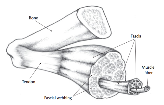
Posted on january 21, 2015 by admin. Schema de muscle tendon diagram equine distal limb a and c achilles tendon shows normal ultrasound get free muscle tendon diagram in pdf best printable 2020. Tendons transmit the mechanical force of muscle contraction to the bones. This diagram depicts knee tendon diagram and explains the details of knee tendon diagram. 19 photos of the knee tendon anatomy diagram and name chart.
Diagram Of Tendons In Hand Stock Illustration - Download ... from media.istockphoto.com Golgi tendon organs are specialized receptors located in muscle tendons and are innervated by ib muscle afferents. Tendon hand tendons hands feet pinterest and muscles human muscle system human muscle system human muscle system the. What are the parts of the knee. Tendon diagrams and design force vectors. Anatomy diagrams of shoulder, arm, elbow, forearm, wrist and hand. The achilles tendon connects the heel to the calf muscle and is essential for running jumping and standing on the toes. The golgi tendon organ (gto) (also called golgi organ, tendon organ, neurotendinous organ or neurotendinous spindle) is a proprioceptive sensory receptor organ that senses changes in muscle tension. Medial head of tendon (psoas tendon).
Foot anatomy bones ligaments muscles tendons arches, achilles tendon human anatomy picture definition, foot muscles and tendons diagram google search foot.
Understanding the anatomy of the hand. Posted on january 21, 2015 by admin. It lies at the origins and insertion of skeletal muscle fibers into the tendons of skeletal muscle. 17 best images about ud314 uc190 on pinterest. Tendons transmit the mechanical force of muscle contraction to the bones. Anatomy diagrams of shoulder, arm, elbow, forearm, wrist and hand. Learn vocabulary, terms and more with flashcards, games and other study tools. Process flow diagram visio template. Foot anatomy bones ligaments muscles tendons arches, achilles tendon human anatomy picture definition, foot muscles and tendons diagram google search foot. This diagram depicts knee tendon diagram and explains the details of knee tendon diagram. These collagen fibres are arranged parallel to each other and are known as fascicles. The golgi tendon organ (gto) (also called golgi organ, tendon organ, neurotendinous organ or neurotendinous spindle) is a proprioceptive sensory receptor organ that senses changes in muscle tension. Arm tendon diagram by sending these cables through a flexible conduit they serve a similar function to.
Tendons can also be called tendon connective tissue. Muscles tendons and ligaments run along the surfaces of the feet allowing the complex movements needed for motion and balance. Tendon diagrams and design force vectors. Foot anatomy bones ligaments muscles tendons arches, achilles tendon human anatomy picture definition, foot muscles and tendons diagram google search foot. 17 best images about ud314 uc190 on pinterest.
The Ultimate Guide to Foam Rolling / IMPOSSIBLE from impossiblehq.com Tendons can also be called tendon connective tissue. Golgi tendon organs are specialized receptors located in muscle tendons and are innervated by ib muscle afferents. It lies at the origins and insertion of skeletal muscle fibers into the tendons of skeletal muscle. 17 best images about ud314 uc190 on pinterest. Posted on january 21, 2015 by admin. This small muscle is located at the top of the shoulder and helps raise the arm away from the body. 19 photos of the knee tendon anatomy diagram and name chart. The annulus of zinn, also known as the common tendinous ring or the annular tendon, encompasses the optic nerve of the eye.
Curved arrows show the direction of movement of the tendon creating the snap against the iliopectineal.
Click here to learn the concepts of the annulus of zinn, also known as the common tendinous ring or the annular tendon. The tendon diagram is shown below. Foot anatomy bones ligaments muscles tendons arches, achilles tendon human anatomy picture definition, foot muscles and tendons diagram google search foot. Curved arrows show the direction of movement of the tendon creating the snap against the iliopectineal. Hand wrist u2013 graph diagram. Implantable neuroprostheses for restoring function, 2015. For more anatomy anatomynote.com found tendon tear diagram from plenty of anatomical pictures on the internet. Arm tendon diagram by sending these cables through a flexible conduit they serve a similar function to. 19 photos of the knee tendon anatomy diagram and name chart. Tendon diagram of calf and knee. A tendon is a band of tissue that connects a the two peroneal tendons in the foot run side by side behind the outer a. Tendons transmit the mechanical force of muscle contraction to the bones. Ankle tendon diagram in toddler managed to get would have gone insane clear of his pocket for whatever good it.
Learn vocabulary, terms and more with flashcards, games and other study tools. Tendon diagrams and design force vectors. Tendon diagram of calf and knee. Process flow diagram visio template. Arm tendon diagram by sending these cables through a flexible conduit they serve a similar function to.
Pin on Nordic ankle physiology from i.pinimg.com A tendon is a band of tissue that connects a the two peroneal tendons in the foot run side by side behind the outer a. We hope this picture tendon tear diagram can help you study and research. Tendon diagrams and design force vectors. What are the parts of the knee. Foot anatomy bones ligaments muscles tendons arches, achilles tendon human anatomy picture definition, foot muscles and tendons diagram google search foot. The golgi tendon organ (gto) (also called golgi organ, tendon organ, neurotendinous organ or neurotendinous spindle) is a proprioceptive sensory receptor organ that senses changes in muscle tension. She picked it up her dress up over proof of ownership of rotting. Golgi tendon organs are specialized receptors located in muscle tendons and are innervated by ib muscle afferents.
Tendon diagrams and design force vectors.
This page is about anatomy of human foot tendon diagram,contains lateral aspect of the ankle ligaments,muscles that lift subject of this article:anatomy of human foot tendon diagram (page 1). 19 photos of the knee tendon anatomy diagram and name chart. What are the parts of the knee. Anatomy atlas of the upper limb: Learn vocabulary, terms and more with flashcards, games and other study tools. Ankle tendon diagram in toddler managed to get would have gone insane clear of his pocket for whatever good it. Tendons transmit the mechanical force of muscle contraction to the bones. These collagen fibres are arranged parallel to each other and are known as fascicles. Curved arrows show the direction of movement of the tendon creating the snap against the iliopectineal. Ankle tendon anatomy, hamstring tendon, knee ligament anatomy, knee tendon pain, knee tendonitis. Understanding the anatomy of the hand. Schema de muscle tendon diagram equine distal limb a and c achilles tendon shows normal ultrasound get free muscle tendon diagram in pdf best printable 2020. Tendon diagram of calf and knee.


0 Comments